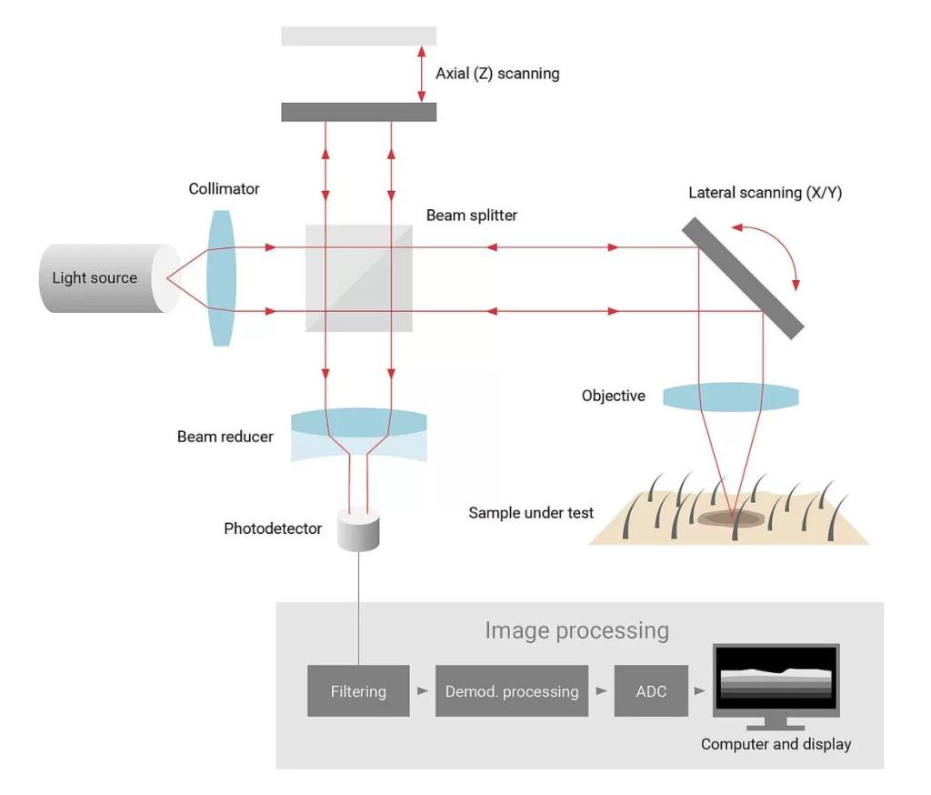
Optical coherence tomography is a powerful imaging technique used to obtain micrometer-scale, cross-sectional images of biological tissues and other scattering media. By using short-coherence-length light sources OCT can discern optical reflections from various depths in the sample. In medical contexts—especially ophthalmology, cardiology, and dermatology—OCT has transformed both diagnostic and therapeutic procedures through its high-resolution, noninvasive imaging capabilities.
At the heart of any OCT system is an interferometric setup, typically a Michelson interferometer with a reference arm and a sample arm. To reconstruct depth profiles (A-scans) at micrometer resolution, the optical path length in the reference arm must be finely tuned to match different layers within the sample. Fiber optic delay lines can provide this precise control allowing one to scan through various depths within the tissue, enabling 2D or even 3D imaging.