What do researchers typically expect from the most cutting-edge microscopes for both live cell imaging and fixed-sample imaging? For example, to image deep within the sample very quickly and sensitively. To image samples of different sizes, from nanometers to millimeters. To be able to discover unforeseen dynamic cellular events. Obtain high-resolution images with nanometer-scale localization accuracy. Imaging living organisms for days at a time without phototoxicity. Obtain as much usable data as possible across the entire field of view.
All this is possible with the new Dragonfly 600 confocal microscope model with dual spinning disk microlenses from ANDOR. The Dragonfly 600 provides excellent multidimensional images from subcellular samples (on the nm scale) to the whole organism (on the cm scale). New super-resolution modules and the patented B-TIRF reveal even the smallest details, such as the dynamics of viral infection or the ultrastructure of chromatin or organelles.
The Dragonfly 600 has all the necessary tools for the single molecule localization method. It uses an astigmatic 3D super-resolution module and provides images with nanometer localization accuracy and a corresponding resolution of up to 10 nm.
This new model builds on the successful Dragonfly 500 series and focuses primarily on DNA-PAINT super-resolution localization imaging and high-resolution 3D tissue imaging. Thanks to its flexible scripting interface, the Dragonfly 600 can be further combined with fluidic systems for large-scale multi-omics imaging.
Where can the Dragonfly 600 be used?
1. Neuroscience
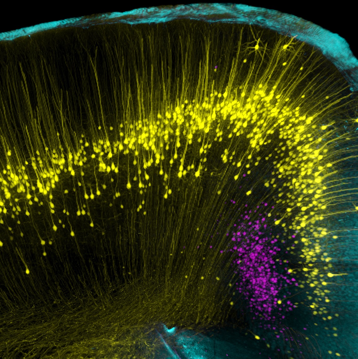
Neuroscience researchers typically need to image both live and fixed samples, in a variety of sizes: from nanometers to millimeters. Almost anything can be imaged with Dragonfly - glial cells, axonal transport, large tissue sections, extended samples (ExM) or dendritic spines, and at super-resolution (SMLM).
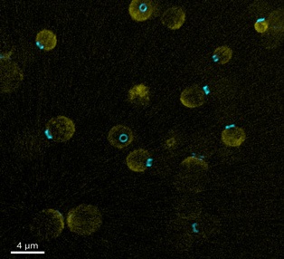
Localizing synaptic zones and understanding functional states using SMLM:
- Super-resolution of synapses in 3D (up to 30 nm axially) using the super-resolution module
- Resolution of bound synaptic vesicles using SMLM with up to 10 nm lateral resolution
- Membrane receptor visibility and signal-to-noise ratio enhancement using B-TIRF
- Super-resolution of structures deep inside neurons using confocal resolution (Z ~ 10 μm)
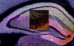
Creating brain atlases using spatial transcriptomics:
- Mapping gene expression at the cellular level
- Accurate quantification of gene expression using Borealis® uniform illumination
- Increase productivity by using large field of view and fast confocal scanning
Imaging of live neuronal samples:
- Imaging dynamic events (e.g. calcium signaling) with fast confocal imaging up to 400 fps
- Imaging of sensitive samples (e.g. mucosal explants, brain sections) with gentle illumination
- Capture all signals with short exposure times and highly sensitive detectors
Imaging deeper into dense brain tissue using NIR:
- Greater penetration into the sample
- Better sample viability due to less energy radiation
2. Cell biology
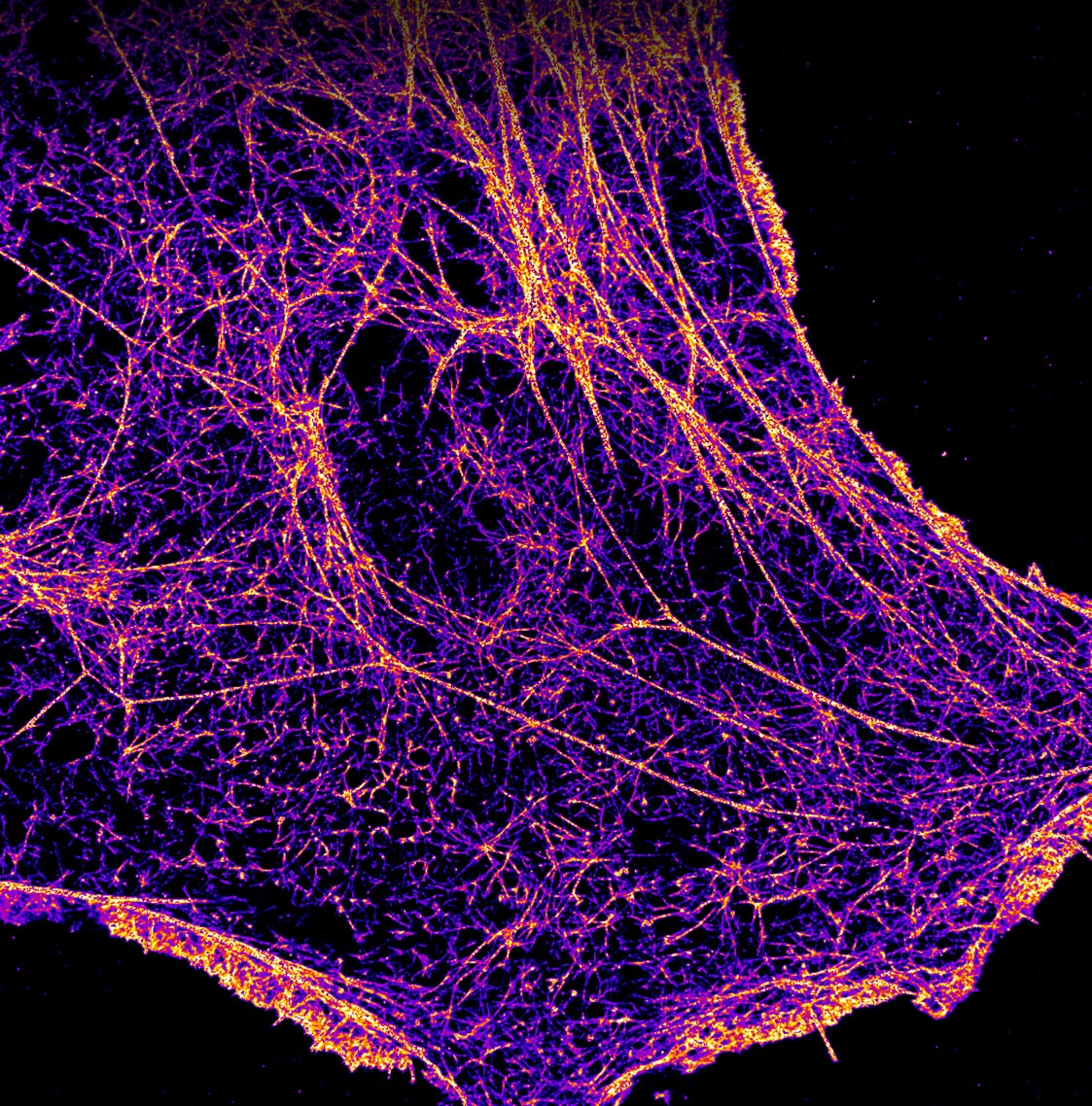
Whether imaging cell populations, intracellular organelles, expansion microscopy (ExM) or super-resolution microscopy, there is always a suitable modality for high-quality cell imaging with Dragonfly 600 - confocal imaging, B-TIRF, widefield, SMLM or super-resolution compatible with live cell imaging (SRRF-Stream+).
Imaging of centriole ultrastructure or 3D nuclear pore complexes:
- Imaging chromatin organization and epigenetics down to 10 nm in XY
- Exploring the structure of the mitochondrial membrane with 30 nm axial resolution using a 3D super-resolution module
- Visualization of receptors on the cell membrane and identification of biomolecules involved in cell signaling using a 3D super-resolution module in combination with HLE and B-TIRF
Imaging cell organelle dynamics with minimal phototoxicity and photobleaching:
- Imaging live samples for several days at a time with highly sensitive detectors
- Imaging of growing microtubules and moving cilia using fast confocal imaging (400 fps)
Imaging of fusion events and receptor signaling
- Gain more information from a single image by uniformly illuminating the entire field of view with patented Borealis technology
- Imaging of vesicular transport using B-TIRF with 30% more uniform illumination than conventional TIRF
Cellular imaging by expansion microscopy (ExM):
- Faster acquisition of multiple images due to large field of view, highly sensitive detectors and high-power lasers
- Quantification and interpretation of more data per microscopic image thanks to Borealis uniform illumination
- Gain information from multiple probes by using the broad excitation spectrum of HLE
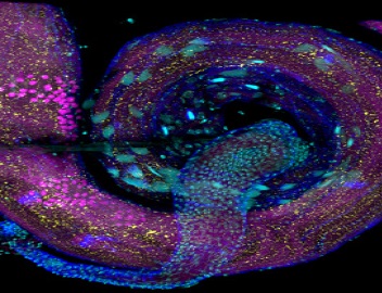
3. Cancer research
"Visualization deep in tissue and in forming metastases thanks to optimized pinhole diameter on the rotating disc."
Cancer research needs a holistic approach to biology requiring insight into gene expression, tumor microenvironment, malignancy and in vitro/in vivo assays. The Dragonfly 600 allows researchers to observe the behavioral interactions of tumor cells with the surrounding environment and the spatial distribution of tumors in fixed or live cell environments.
With the broad excitation spectrum of ILE or HLE and multi-color imaging of multiple tumor signals, it is possible to capture all information in a single experiment. Sparing imaging provides live images of tumor tissue over hours or days. The speed of imaging and optimized pinhole spacing on the spinning disk allows for more in-depth imaging of organoids and dense tissues.
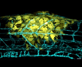
4. Developmental biology
"Imaging blood flow or cilia movement using confocal imaging."
Developmental biology requires imaging of thicker specimens. The optimized size and spacing of the pinholes on the spinning disk of the Dragonfly system allows imaging of thicker samples down to millimeter depth. With uniform illumination of the entire field of view without edge vignetting, multiple images can be taken at high magnification, seamlessly stitched together as if they were a single image, and the entire organism can be visualized at high resolution. Thanks to the exceptional speed of the Dragonfly system and the highly sensitive detectors, e.g. the development of mouse embryos or other challenging processes can be imaged for hours or days.
5. Microbiology and virology
"Visualizing host-pathogen interactions with the increased uniformity provided by the new B-TIRF."
With the unique B-TIRF module on the Dragonfly 600, a vivid view of virus-host interactions can be obtained. By combining a powerful HLE laser engine with a 3D super-resolution module that collects data with up to 10 nm lateral resolution and 30 nm axial resolution, information on the ultrastructural biology of fungi, bacteria and viruses can be obtained. Taking advantage of the high background suppression and extreme sensitivity of the detectors, infection deep within tissues can be analyzed.
6. Transcriptomics & proteomics
"Increased productivity in omics data acquisition with a high-power HLE laser engine."
Understanding the molecular basis of development, brain function, neurodegenerative diseases and their behavior is a huge task. The scientific field known as "-omics" gathers information on Xn biological molecules to characterize and quantify the entire set of molecules. The Dragonfly system is ideally suited for such applications, thanks to its highly sensitive detectors, extremely high background suppression, scanning speed, uniform illumination and automation capabilities. Research in -omics has applications in many areas of life sciences, such as predicting disease progression based on gene expression maps, access to the tumor microenvironment , and cancer severity.