The FLIM (Fluorescence Lifetime Imaging Microscopy) method uses an optical microscope to image the lifetime of fluorescence. It is a fluorescence microscopy technique that uses fluorescence to create contrast in individual microscopic images. The output is a so-called FLIM map, which gives the spatial distribution of the fluorescence lifetime for a selected region of the sample. The FLIM setup can be implemented by combining an optical microscope with a TCSPC luminescence spectrometer.
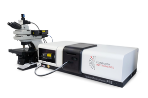
Standard configuration of the fluorimeter plus microscope assembly
Fluorescence broadband imaging
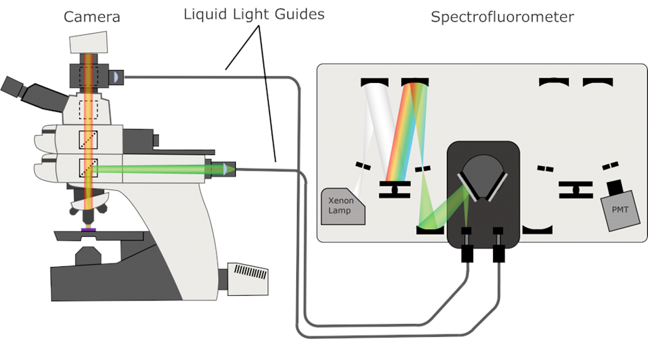
In this configuration, a broad-spectrum Xe lamp is used to excite the sample, which is part of the luminescence spectrometer. An excitation monochromator is used to select the desired wavelength, the output of which is fed to the microscope via a liquid light guide or optical fibre. The excitation beam is reflected from a dichroic mirror inside the microscope and directed into the objective. A sensitive camera is used to record the luminescence image.
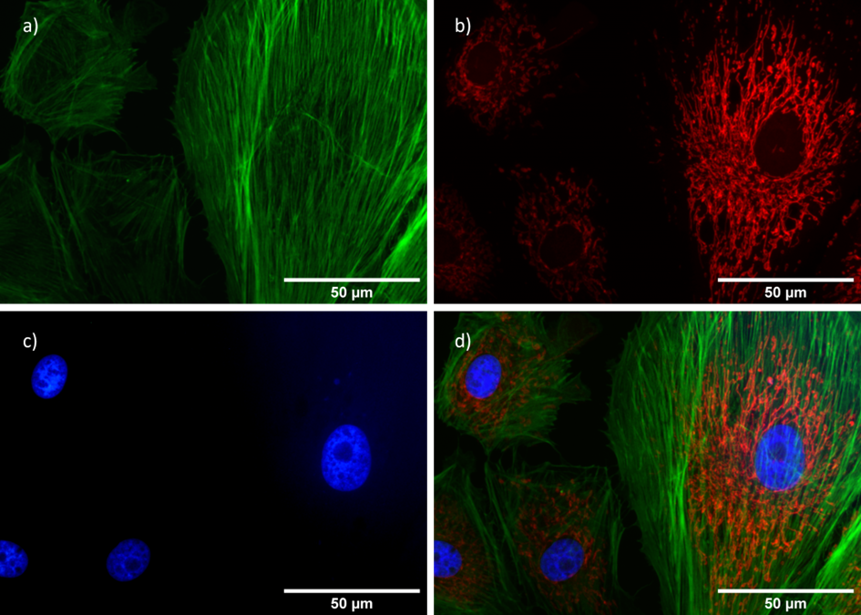
- broadband fluorescence image of BPAE (bovine pulmonary artery endothelial) cells
Detection of luminescence spectra
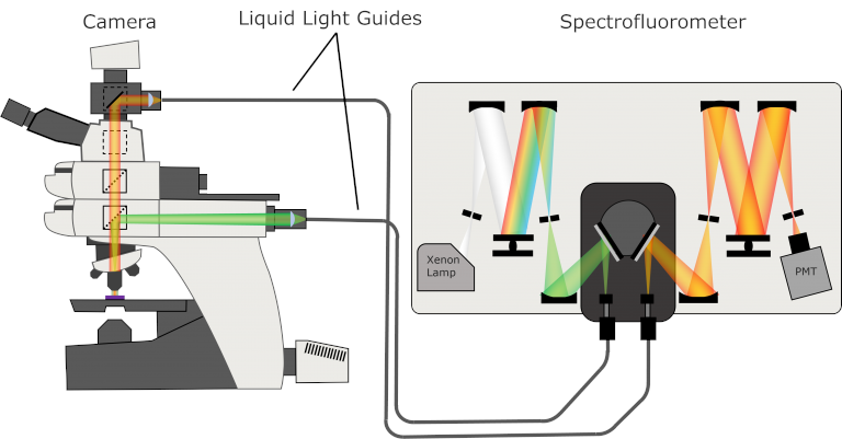
Both the excitation and emission branches of the spectrofluorimeter are used in this setup. Liquid light guides (optical fibres) provide the excitation signal from the fluorimeter to the microscope and the luminescence signal from the microscope to the fluorimeter.
Fluorescence/phosphorescence lifetime detection
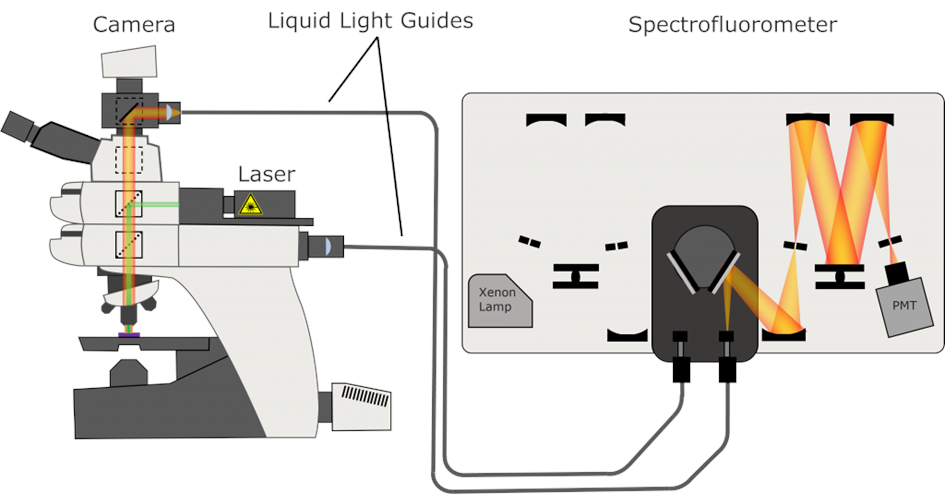
To measure the lifetime of the luminescence, a pulsed laser diode is integrated into the microscope as an excitation source. The laser beam is directed to the desired location on the sample using a camera and a sliding stage. The luminescence signal is fed into the emission arm of the TCSPC spectrometer by means of a liquid light guide (or optical fibre).
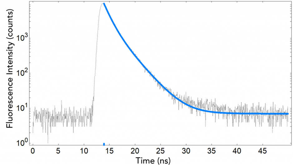
- DAPI fluorescence decay after binding to DNA, lifetime determined by TCSPC technique
FLIM (Fluorescence Lifetime Imaging Microscopy)
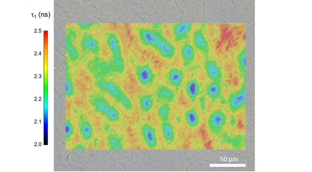
If the microscope includes a computer-controlled XYZ stage, then the entire assembly can be used in a Fluorescence Lifetime Imaging Microscopy (FLIM) configuration to detect luminescence lifetimes. In this fluorescence imaging method, the contrast of the resulting image is based on the lifetime of the individual fluorophores. The resulting map gives the spatial distribution of the fluorescence lifetime for a selected region of the sample. In addition to pulsed laser diodes, supercontinuum lasers, which generate pulsed (picosecond) broad-spectrum radiation, are increasingly used to excite luminescence. This gives the user much more flexibility in the choice of excitation wavelength.