

| Delay lines | |||||||
|---|---|---|---|---|---|---|---|
| Operating wavelength [nm] | 1060 | 1550 | 2000 | ||||
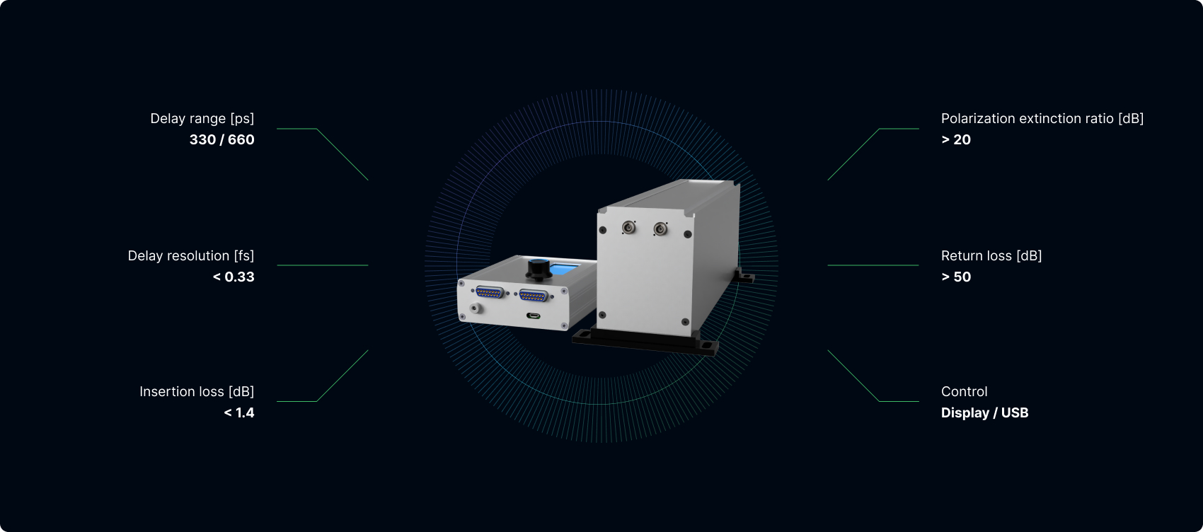
| Optical delay range [ps] | 330 | 660 | 330 | 660 | 330 | 660 | |
| Typical insertion loss [dB] | 1.4 | 1.7 | 1.4 | 1.7 | 2.5 | 2.5 | |
| Insertion loss uniformity [dB] | 0.25 | 0.35 | 0.25 | 0.35 | 0.2 | 0.2 | |
| Optical delay resolution [fs] | < 0.33 | ||||||
| Polarization extinction ratio [dB] | > 20 | ||||||
| Fiber type | PM-980-XP / 1060-XP | PM-1550 / SM-1550 | PM-1950 / SM-1950 | ||||
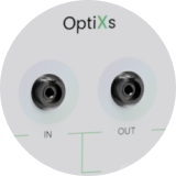
| Fiber connectors | FC/APC, FC/PC | ||||||
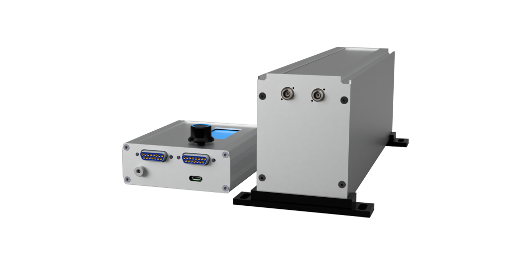
| Dimensions (delay line) [mm] | 76.4 x 106 x 231.5 / 76.4 x 106 x 286.5 | ||||||

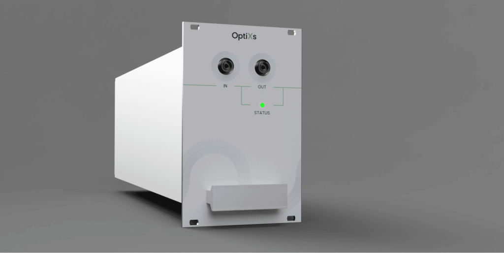
Designed by OptiXs
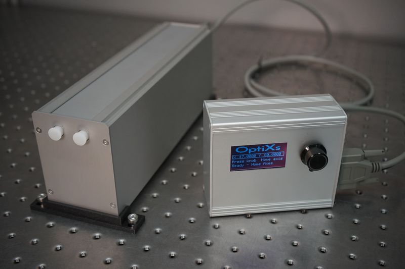
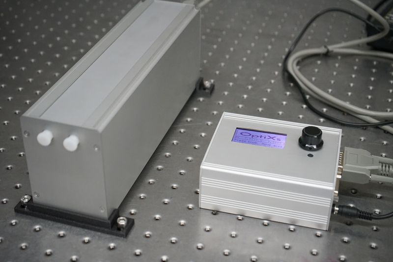
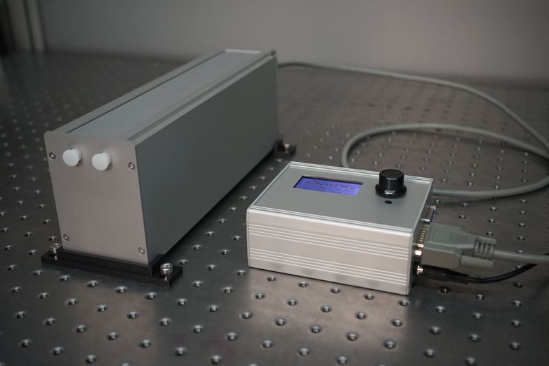
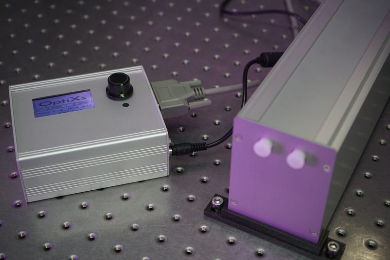
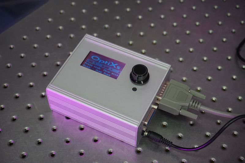
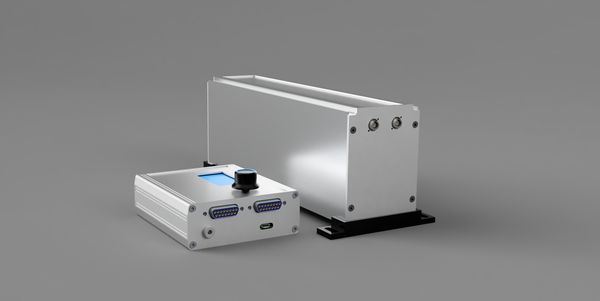
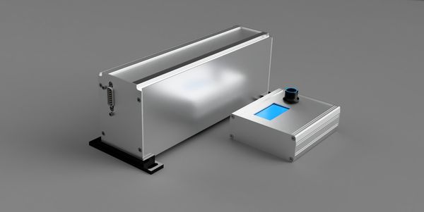
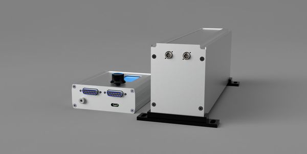
FODL-SA
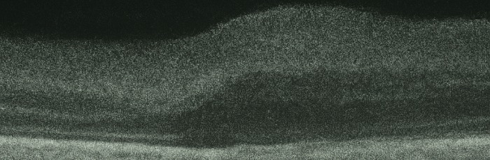
Fiber delay line with optical delay range up to 660 ps and resolution < 0.33 fs. Controller with display and USB connection for remote control. Ideal for OCT, TDM, research and industrial applications.
Send inquiry