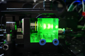
SPIM, or thin-sheet illumination microscopy, is a non-invasive method of wide-field microscopy based on illuminating a sample with a laser beam stretched by a cylindrical lens or vertical aperture into a line shape (the method is then called light sheet microscopy). Due to the perpendicular positioning of the camera (detector) to the laser beam, the image is projected from the focal plane onto the detector in a 2D projection. This arrangement contributes to the suppression of noise (arising outside the focal plane in the form of a signal) and to the production of images approaching the quality of images from a confocal microscope. By moving the sample, optical sections can be obtained from which a 3D reconstruction of the image can then be created. By filming the sample and taking images at different angles, a so-called multi-view 3D model can be obtained by subsequent digital reconstruction, which contains high-resolution information about the entire sample.
SPIM is one of the ideal methods for live cell imaging due to its unique properties (limited scanning area, low incident light intensity, high scanning speed limited only by the camera scanning speed).


