We distribute this product only in the Czech Republic and Slovakia.

Andor BC43 CF compact confocal microscope

Andor BC43 confocal microscope

Small in size, big in power - that's the new plug & play confocal microscope from ANDOR, suitable for both beginners and experienced microscopists!

Thanks to its compact dimensions (505 x 633 x 443 mm - W x D x H), the BC43 CF fits into even a small working space, and also benefits from a sophisticated design with a lightweight, tight and opaque cover and a built-in anti-vibration plate, so there is no need for a darkroom or separate optical table.

Less time for training, more time for imaging!

The BC43 CF is an intuitive microscope that even novice scientists can easily master thanks to its simple operating procedures and minimal maintenance!

Alexandra Pešátová
Expert advisor

Mgr. Alexandra Pešátová

+420 725 945 076

pesatova@optixs.cz

Send inquiry

OptiXs care

  • We will expertly consult your planned application
  • Our team is able to integrate the product into a larger system
  • We ensure fast delivery of spare parts and local service
What else we can help with

Popis produktu

Andor BC43 confocal microscope

Small in size, big in power - that's the new plug & play confocal microscope from ANDOR, suitable for both beginners and experienced microscopists!

Thanks to its compact dimensions (505 x 633 x 443 mm - W x D x H), the BC43 CF fits into even a small working space, and also benefits from a sophisticated design with a lightweight, tight and opaque cover and a built-in anti-vibration plate, so there is no need for a darkroom or separate optical table.

Less time for training, more time for imaging!

The BC43 CF is an intuitive microscope that even novice scientists can easily master thanks to its simple operating procedures and minimal maintenance!

Accessories

Integrated software

Fusion software

Complementary software for control and basic image data processing - FUSION- is integrated into the BC43 CF, providing superior imaging in a few easy steps. The advantage of the Fusion software is primarily:

  • Real-time 3D data visualization simultaneously with scanning
  • User-friendly interface with intuitive controls
  • Easy setup of protocols for multidimensional experiments
  • Post-acquisition data processing - GPU-accelerated stitching and deconvolution(ClearView-GPU)
  • Automatic image capture of large samples (larger than the field of view) and stitching into 2D and 3D montages for a complete sample image
  • Patented Focus Seek & Lock autofocus technology - to simplify focusing on samples and maintaining focus during long time-lapse shots and acquisition of large samples
Imaris

Also at the heart of the BC43 CF is the core software package for visualization and analysis of microscopy data - IMARIS, which builds on Fusion's common *.ims data format to speed and simplify the transition between image data acquisition and analysis. Imaris software allows users to:

  • Visualize their microscopy images in 2D/3D/4D quickly and easily.
  • Create iso-surface reconstructions for better interpretation and presentation of raw data
  • Easily create high-resolution images and multi-dimensional video sequences

Additional application-specific modules can be added to the basic Imaris package to extend the analysis capabilities, e.g. Imaris for cell biology, neuroscience, etc.

Mammalian cell

Mammalian cell in prophase. Image was acquired using BC43 confocal mode, using 4 acquisition channels and covering 10 μm Z range at Nyquist. Image was further deconvolved and rendered in Imaris. Dark blue - actin, yellow - microtubules, magenta -mitochondria, cyan-DNA. Image credits: Claudia Florindo, Andor Technology.

Video

Key features of BC43 CF

Hardware:

  • High-speed confocal imaging to capture dynamic events in thicker samples
  • Widefield mode for light-sensitive samples and to detect the weakest signals
  • Flexibility in imaging, ability to combine multiple imaging modalities in a single protocol
  • Desktop compact device with opaque, dark enclosure (eliminates the need for a dark room) and built-in anti-vibration mechanism
  • 5-position motorized lens carousel
  • Efficient navigation, position and focus per sample with adjustable navigation and focus speed via ergonomic 3D joystick
  • Patented Borealisillumination system for confocal and widefield imaging providing uniform illumination across the entire field of view and greater light beam transmission
  • Highly sensitive detector for short exposures and photobleaching reduction, offering high dynamic range to capture both weak and bright signals in a single frame without saturation
BC43 - jednotlivé části

Software:

  • Complementary Fusion control software with ClearView-GPU deconvolution module (up to 50x acceleration via GPU) to provide increased image resolution and contrast and improve overall image quality
  • Simple workflow from sample insertion to image acquisition - just insert the sample, find the point of interest, set the thresholds, and get the image with a single click. No need for lengthy training or previous experience.
  • Patented Focus Seek & Lock technology for improved sample focusing even in longer time-lapse experiments
  • Multi-dimensional display function for visualizing the entire sample - simultaneous creation of time-lapse images, Z-stacks and tile positions
  • Automatically acquires data of large samples (larger than the field of view) and stitches them into 2D and 3D montages for a complete image
  • Enables multiwell imaging - displays 6, 12, 24 and 96-well culture plates
  • Real-time3D dataimaging provides instant visual feedback on the progress of the experiment for real-time data evaluation and visualization
  • Basic Imarissoftware package for visualization and comprehensive analysis of hundreds of GB of 2D/3D/4D microscopy data, expandable with application-specific modules
Zebrafish

Zebrafish fin in the process of bone regeneration. Image shows the perfect stitching of 4 imaging fields, using three channels and 51 stacks for each field, covering a Z range of 174 μm. Newly formed bony tissue in purple (calcein staining) and cathepsin k+ cells (the osteoclasts) in yellow, DNA is in Cyan. Image credits: Alessio Carletti, Universidade do Algarve

Andor BC43 CF confocal microscope specifications

The main microscopic unit:

  • Imaging modes: spinning disk confocal microscopy, epifluorescence widefield, transmitted light - brightfield and differential phase contrast
  • Imaging methods: single color, multicolor, Z-stacks, time-lapse, multiposition, multiwell, montage and 2D/3D stitching
  • ClearView GPU: removes non-specific background signals in the sample and enhances resolution beyond normal optical limits

Camera:

  • Resolution: 6.5 μm pixel; 2048 x 2000 pixel (4.1 MPx)
  • Quantum efficiency: up to 82%
  • FOV: 18.4 mm diagonally
  • Cooling: 0°C (32°F)
  • Images: monochrome, 16bit

Illumination:

  • Fluorescence source: 4 fixed wavelengths 405 nm, 488 nm, 561 nm, 638 nm
  • Transmitted light: broad spectrum LED

Optics:

  • Motorized carousel for 5 lenses
  • Lens magnification: the microscope always includes a 2x magnification objective to facilitate sample navigation. The remaining 4 positions can be fitted with lenses of your choice and requirements
  • Emission filter set for imaging commonly used fluorophores (DAPI, Alexa 488/GFP, Alexa 546/mCherry/TRITC, Cy5/Alexa Fluor 640)
  • Motorised XY stage: positioning range 110 x 80 mm; resolution 100 nm
  • Z-control & Focus: 14.5 mm positioning range
  • Seek & Lock Technology: automatically find the focal plane and maintain focus stability during time-lapse experiments
  • Sample holders: standard slides (25 x 75 mm), culture dishes (35 mm diameter), multiwell culture plates (6, 12, 24 and 96), cover slips for multiwell chambers (2, 4, 8)
  • Incubator (optional item): Stage-top, sliding lid for easy access and sample change, heater for oil immersion lenses

Workstation:

  • PC: Windows™ 10 software, 64 GB DDR4 RAM, 512 GB PCIe SSD Boot drive, 4 GB graphics card, 2 TB image data storage

Flatfish image acquired with Andor Bench Top Confocal BC 43 using multiple tile acquisition and montage. 6 tiles were acquired to compose the image covering a range of 554 µm. The image was rendered with Imaris. Image credit: Marco Campinho, University of Algarve and Claudia Florindo, Andor Technology.

Drosophila egg chamber. The image is a maximum intensity projection of 309 Z planes overing a range of 93 µm. The image was deconvolved using ClearView and rendered in Imaris. (Yellow-DNA, Cyan-actin). Image credit: Rui Silva, University of Algarve and Claudia Florindo, Andor Technology.

This uninjured adult digit (12 week old mouse) comes from a transgenic animal Dmp1 Cre ERT crossed to a TdTomato reporter. The magenta signal correlates with the Td Tomato reporter which would be marking cells of the bone linage (Dmp1 positive) and the 'cyan' is a simple Hoechst stain. Image credit: Dr Mekayla Storer, SickKids, Toronto, Canada.

Confocal Imaging modality of the Andor Benchtop confocal BC43 was used to acquire an early-stage Drosophila Embryo. The image presented from this experiment is a result of the stitching of 6 imaging fields, over a Z range of 40 µm. Microtubules are labelled blue, Centrosomes in Yellow and DNA as Magenta. Image credit: Ines Baião-Santos and Álvaro Tavares, University of Algarve, Claudia Florindo, Andor Technology.

GFP and mCherry labeled membrane markers expressed in Drosophila salivary gland cells. Image credit: Cheng-I Jonathan Ma and Julie Brill, Cell Biology Program, The Hospital for Sick Children.

Development of a Drosophila embryo is effortlessly visualised in 3D with up to 4 channels using the confocal imaging mode of the BC43. Image credit: Ines Baião-Santos and Álvaro Tavares, University of Algarve, Claudia Florindo, Andor Technology.

Chicken embryo stage HH15/16 stained with DAPI. At this stage of development, the notochord, neural tube and somites are clearly visible. Image credit: Tomás Azevedo, University of Algarve and Claudia Florindo, Andor Technology

Drosophila embryo stained with neuronal marker were set up on a multi-position/montage acquisition. This selected image is a montage of a 3X3 field at the selected position, with 101 z slices covering 50 µm range. Image credit: Álvaro Tavares, University of Algarve and Claudia Florindo, Andor Technology.

This image is a single 3D view from a timelapse experiment - cell division can clearly be observed throughout the developing Drosophila embryo. DNA is in mangenta, microtubules are in Cyan. Image credit: Ines Baião-Santos and Álvaro Tavares, University of Algarve, Claudia Florindo, Andor Technology.

Application

Parameters

Confocal microscopy, Widefield, Brightfield, DIC - just all in one!

The Andor BC43 CF(Andor Benchtop Confocal) uses the SDCM imaging technology of the Dragonfly system to offer absolute flexibility in imaging - at the touch of a button, it provides high quality and fast 2D and 3D routine imaging from brightfield to confocal microscopy regardless of sample type.

1. Confocal mode

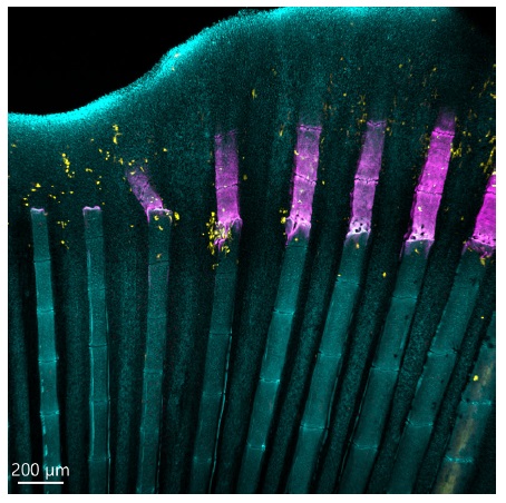
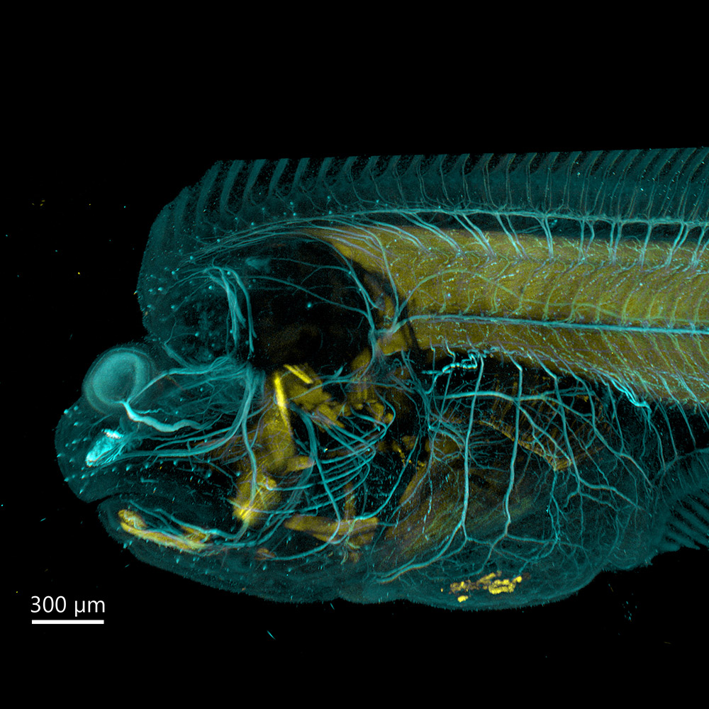
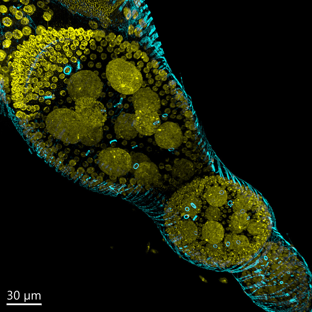
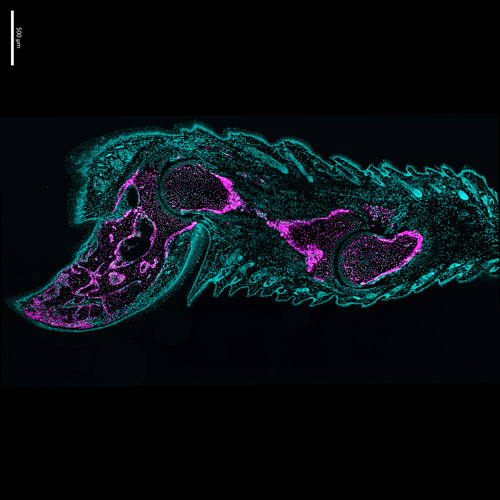
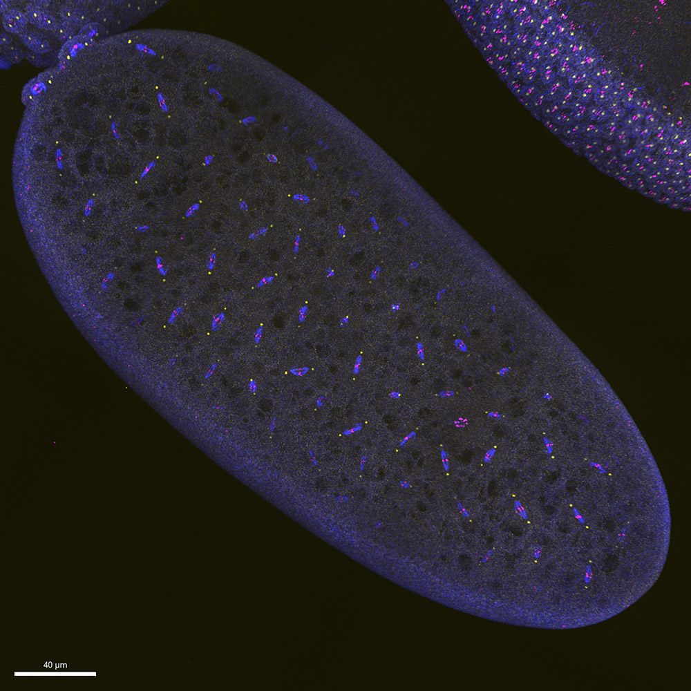
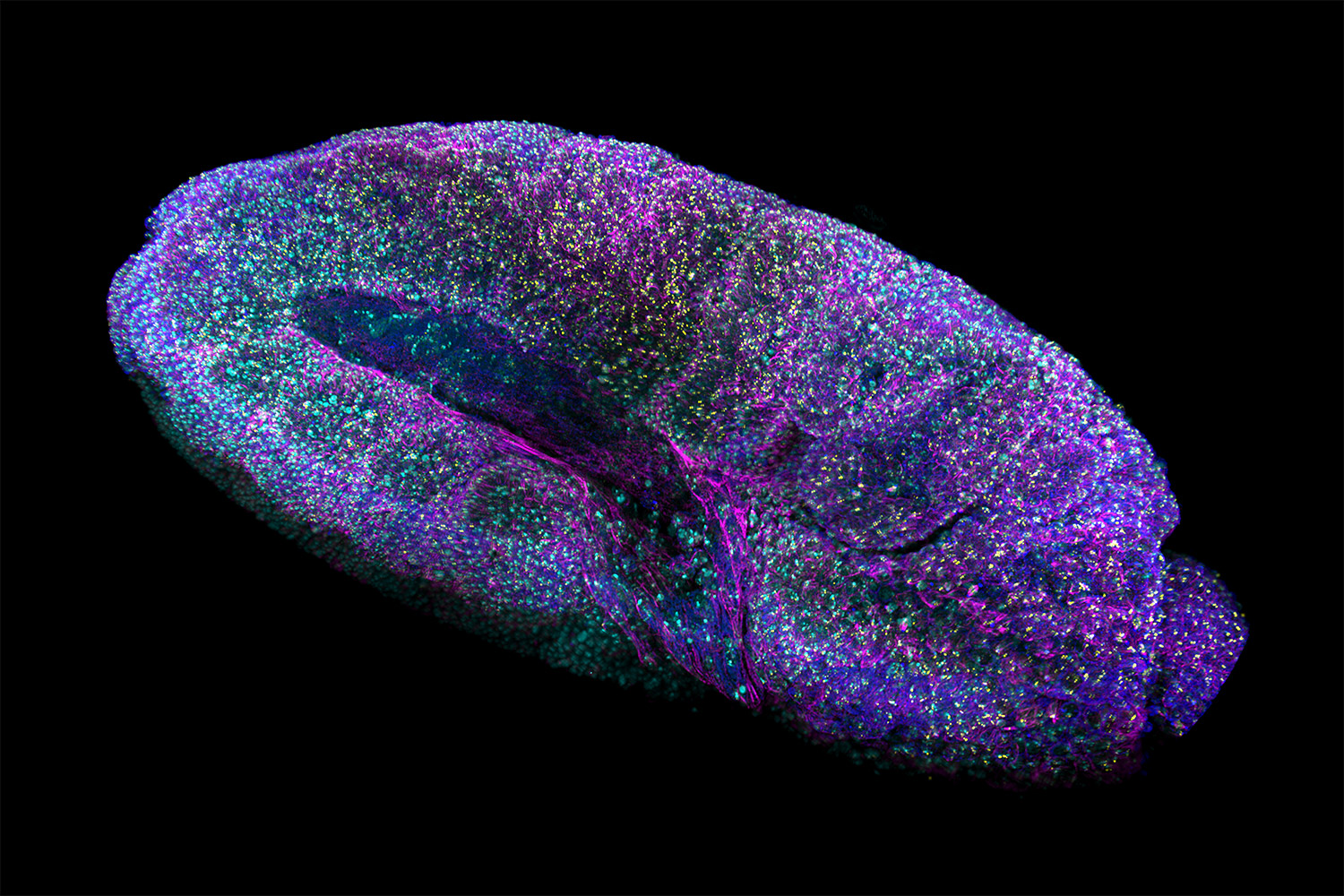
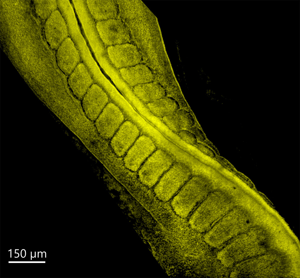
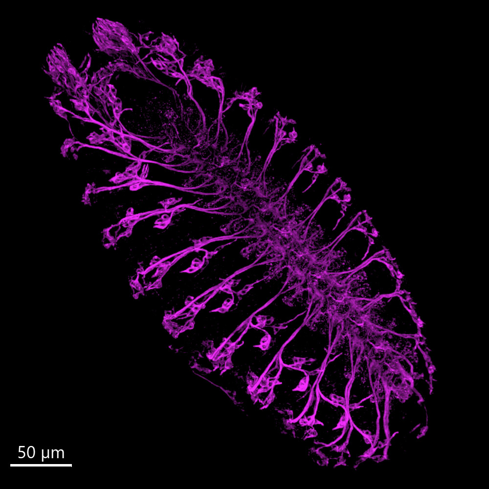
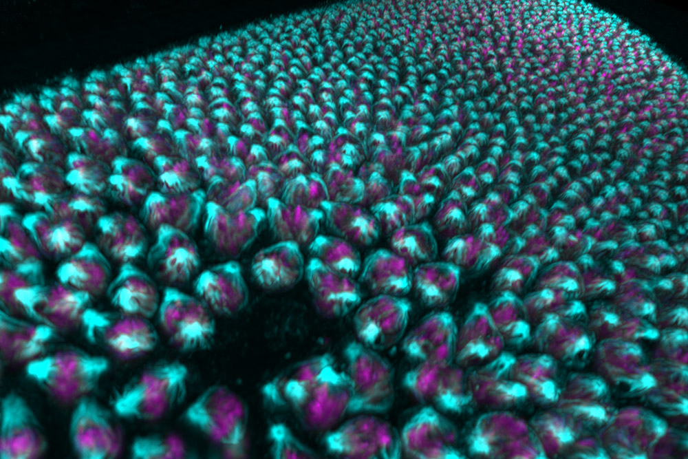
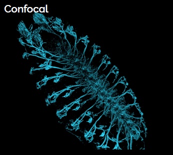
The confocal imaging technology provides high contrast and sharpness, which not only enhances the image quality of thin samples such as monolayer cultures, but is particularly suitable for thicker samples (small organisms, 3D cultures, tissue samples, etc.).

Confocal mode

Compared to point scanning confocal microscopes, the Andor BC43 CF captures images more than 10 times faster while maintaining full resolution. Like the Dragonfly system, the new BC43 CF microscope is based on a spinning disk (a pair of "pinhole disks" with pinholes and a "collector disk" with microlenses). The high-sensitivity, high-speed sCMOS detector for short exposures and photobleaching reduction is the ideal choice for confocal imaging of live cells, maximizing the number of cells per image and efficiently capturing even large samples with a large field of view. Thanks to its high dynamic range, it captures weak and bright signals in a single image without saturation.

2. Widefield mode

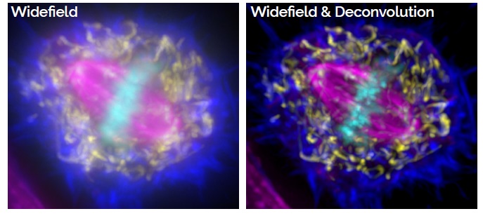
Offers the most user-friendly illumination mode for samples that are super-sensitive to light. It allows detection of even weaker fluorophore signals than in confocal mode. Provides the highest imaging speeds for capturing dynamic processes in living cells. Imaging of thin samples and structures that do not require optical sectioning.

Widefield

In addition, it eliminates sample blurring with a dedicatedClearView-GPU deconvolution module (accelerated via GPU up to 50x) to increase image resolution - ensuring the best image quality and clarity!

3. Passing Light Mode

DPC + konfokál

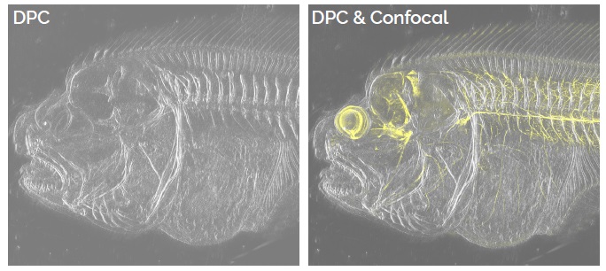
The BC43 CF provides two different options for transmitted light imaging - brightfield mode for visualizing the development of larger organisms whose structures naturally provide high contrast images, and differential phase contrast (DPC) mode for visualizing and rendering the structure of thinner and/or higher transparency samples.

The different modes can be combined for even greater imaging flexibility!

Referenční projekty

Dokumenty

Kompaktní konfokální mikroskop Andor BC43

Inquire product

Are you interested in product? Send us your requirements via the enquiry form or contact an expert consultant directly. We will be happy to answer your questions and propose a solution according to your needs.

Expert advisor

Alexandra Pešátová

Mgr. Alexandra Pešátová