Live cell microscopy(live cell imaging) is a very popular method in the field of cell biology. Obtaining correct and high quality information is subject to several specific factors that can significantly affect live cell experiments. For live cell imaging, we recommend a spinning disk confocal microscope ( SDCM ) or a widefield fluorescence microscope with deconvolution software.
In live cellimaging, great care must be taken to reduce the photo-toxic effects of light(bleaching). The illumination power density at the sample plane, compatible with the life of the cells under investigation, ranges from mW/cm2(widefield fluorescence microscopes) to W/cm2( spinning discconfocal microscopes SDCM ). At high light power densities, intense bleaching of fluorophores occurs, generating free radicals and other reaction products that destroy surrounding molecules and alter the environment outside and inside the cells.
Cell cultures and tissues must be maintained under physiological conditions throughout the experiment. Factors such as temperature, humidity, medium composition and pH are very important to obtain meaningful information about the processes under investigation. Depending on the origin of the cultured cells, the above factors need to be set to specific values and kept stable.
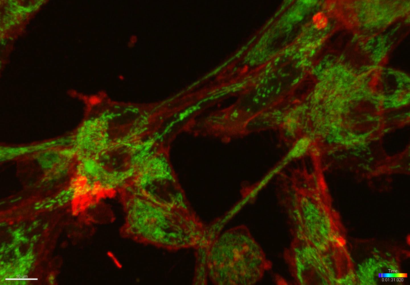
Mitochondria in glioma cells. iXon Ultra, 60x, NA: 1.49. A total of 6,478 1Mpx images were taken! Voxel 0.201*0.201*0.293 um, X/Y:1024*1024 pixels, Z:41, T:79, 2 channels. The data was taken during a short (13 min) time-lapse experiment during the demonstration of the Andor Dragonfly system. Samples were provided by Dr. Veronika Huntošová from UPJS/CIB, Košice, Slovakia.
Essential equipment for live cell imaging microscope is listed in the table:
| Equipment | Contribution | Reason | Manufacturer |
|---|---|---|---|
| Confocal microscope (SDCM) |
Low bleaching High scanning speed |
Pinhole kit disk ensures confocality Illuminates the entire field of view at once Use of cameras for emission signal detection |
Andor |
| Deconvolution |
3D reconstruction of widefield fluorescence microscope images Higher resolution |
Algorithms for removing signal from out-of-focus planes Signal reorganization according to the parameters of the microscope used |
Bitplane |
| Cameras |
High sensitivity High speed High resolution |
Sophisticated sensor architecture (sCMOS/EMCCD) Fast signal readout |
Andor |
| Incubator | Temperature, humidity, pH control | Isolation of the sample from the surrounding environment and precise control of the conditions during the experiment | Okolab |
| Motorized stage |
High resolution Repeatability |
Precision compontents Piezo motors for maximum resolution |
PI |
| Visualization and analysis |
3D visualization Analysis of acquired information Comparison of results |
Colocalization (2D/3D colocalization) Tracking (2D/3D tracking) Statistical data acquisition Creation of graphs |
Bitplane |
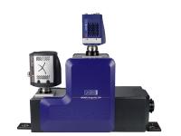
The Andor Dragonfly confocal microscope meets all the requirements for live cellimaging:
Scanning speed
- 400 frames per second (512 × 512 pixels)
- Minimum exposure time - 2.5 ms
Sensitivity
- 90% quantum efficiency for iXon cameras (EMCCD)
- 82% quantum efficiency for Zyla cameras (sCMOS)
Environmental control
- Incubators from world leading manufacturers (Okolab)
Microscope
- Precision XY and Z piezo motorized stage
- Hardware autofocus
- Specialized lenses
More
- Active blanking - connection between light source and cameras
- Filters - compatible with required wavelengths in excitation and emission paths