The Raman effect is the inelastic scattering of light on particles (molecules and atoms) in which the particles transition to one of the quantum states by transferring a quantum of energy. Raman spectroscopy is a very efficient and non-destructive technique that provides information about vibrational and rotational transitions in molecules. It is used in many applications, including basic research, routine process control and materials identification.
Raman scattering spectra (Stokes and anti-Stokes branches)
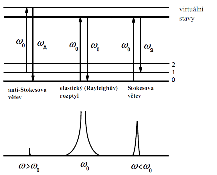
A molecule first absorbs a photon of excitation radiation (is excited from an initial state to a virtual energy state). On relaxation, the molecule then emits a photon and enters a final (vibrational or rotational) state that is not identical to the initial energy state of the molecule. The energy difference between the initial and final states leads to a shift in the frequency of the emitted photon away from the excitation frequency ω0. If the final vibrational state has a higher (or lower) energy value than the initial state, then the emitted photon will be shifted towards lower (or higher) frequencies. Thus, the Raman scattering spectrum consists of pairs of lines that are symmetrically distributed with respect to a line of elastically scattered radiation of frequency ω0 (see figure). The region of lower frequencies is called the Stokes branch, and the region of higher frequencies is called the anti-Stokes branch of the Raman spectrum. In practice, the spectrum of the Stokes branch of the Raman spectrum is usually measured as it has a much higher intensity.
Special techniques of Raman spectroscopy
The Raman scattering spectrum of a substance is very weak in intensity, which makes many measurements uncomfortable. This is one of the reasons why special Raman spectroscopy techniques that can overcome some of the limitations of the standard Raman phenomenon are becoming increasingly popular.
Resonance Raman spectroscopy
If the wavelength of the excitation laser used corresponds to the energy required to move an electron in the molecule of the substance under investigation to a higher energy level, the resonant Raman effect can be amplified by 2 to 4 orders of magnitude. Each type of bonding of a molecule corresponds to a different wavelength. We then speak of a selective resonant Raman effect.
Surface-enhanced Raman spectroscopy
Surface-Enhanced Raman Scattering (SERS) refers to the significant enhancement of Raman scattering (typically103 to106) due to the interaction of visible radiation with plasmonic metal nanostructures and with molecules localized on their surfaces. The molecules under investigation are first absorbed on a suitable metal substrate. SERS substrates containing gold or silver nanoparticles are most commonly used. Two mechanisms contribute to the overall enhancement of the Raman scattering intensity: an electromagnetic effect (resonant excitation of surface plasmons in the metal) and a chemical effect (charge transfer between the absorbate and the metal surface).
Time-resolved Raman spectroscopy
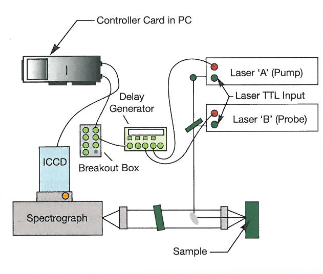
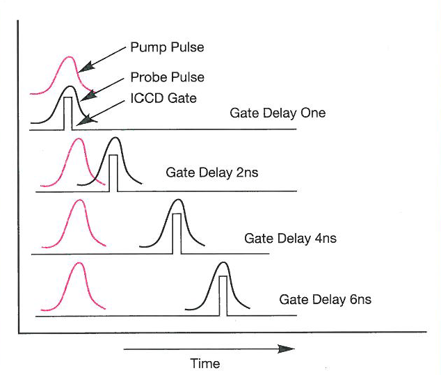
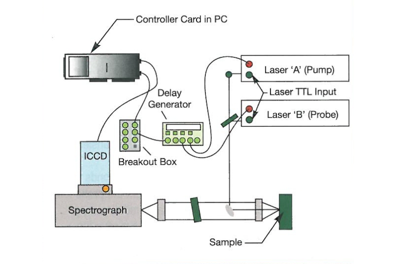
Time-Resolved Raman Spectroscopy (TRRS) makes it possible to study microscopic processes that take place over very short time intervals. The incident laser pulse brings the material under investigation out of thermodynamic equilibrium and the individual molecules are in an excited state. The same laser pulse (single-colour pulsed Raman experiment) or a pulse originating from a different laser source (two-colour pulsed Raman) is then used to obtain the Raman spectrum of the excited molecules. The attached figure shows a typical experimental setup for two-color time-resolved Raman scattering measurements using two lasers, a spectrograph with an ICCD camera and a delay generator. One laser is used to excite the sample to be measured, the other is used to probe the changes induced in the substance by the excitation pulse. The time course of the transient signal is monitored by recording a series of spectra corresponding to different moments after photoexcitation.
Raman microscopy
Raman microscopy combines Raman spectroscopy with microscopic methods. This approach undoubtedly has many advantages and brings with it new possibilities for sample identification. A typical example isTip-Enhanced RamanSpectroscopy (TERS), which uses Raman spectroscopy in conjunction with atomic force microscopy with a special AFM tip to extract information from structures at the nanometer level.