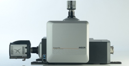

The Dragonfly 200 confocal microscope is the ideal solution for visualizing living cells or organisms - live cell imaging Live cell microscopy, or live cell imaging, is a popular method in the field of cell biology.
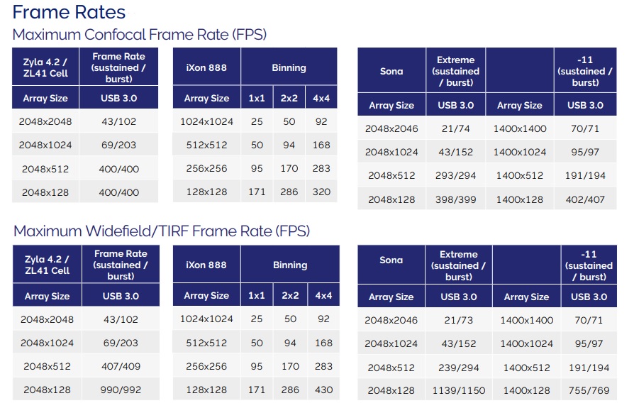
Due to the high scanning speed and extreme sensitivity of the detectors, the toxic effects of light and bleaching fluorophores are reduced. The acquisition time for a high-quality image is minimal compared to scanning confocal microscopes (LSCM), which is an advantage not only when fast processes need to be captured, but also for routine spatial imaging of fixed samples.
Key features
- Fast and gentle confocal microscope - Spinning disk
- Resolution of 2048 × 2048pixels and more - SRRF Stream
- Wide field imaging by extending the disk - Widefield
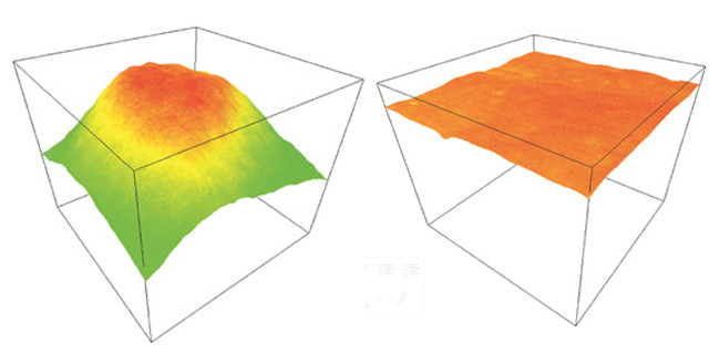
- Integrated Borealis sample perfect illumination system
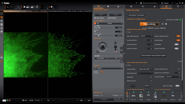
- Complementary software with deconvolution and super-resolution- Fusion
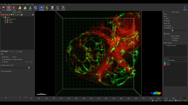
- Image analysis and 3D visualizationsoftware - Imaris Core
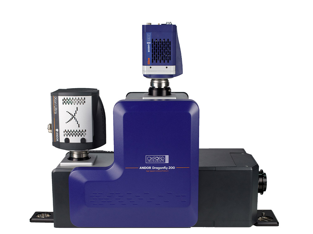
Unparalleled speed and sensitivity of imaging is due to the use of state-of-the-art high quantum efficiency camerasfrom Andorand the patented Borealis illumination system. The Dragonflyconfocal microscope is complemented by the new, complementary Fusion software. Also included is the basic version of Bitplane's excellent image analysis and 3D visualization software, Imaris Core.
The Dragonfly 200 confocal microscopeprovides even more flexibilityregarding the microscope body. Both direct and inverted microscopesfrom the world's leading manufacturers are compatible. The technology behind the excellent performance is identical for both systems. For detailed information on the differences in configuration, please refer to the technical specification below.